
by Alan Jordan
Modic changes as seen on MRI are an inflammation/infection in the vertebral body adjacent to the disc – either above/below or both – in patients that have experienced a disc herniation. In some instances the cause of the inflammation is bacterial while in others it is likely to be of mechanical origin. Examples of Modic changes types 1 and 2 can be seen below. These are the most closely associated with lower back pain.
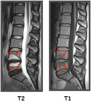
Modic Changes type 1 as seen on two different MRI scan settings.
Modic Changes type 2 as seen on two different MRI scan settings.
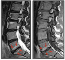
What we see here are inflammatory changes in the vertebral bodies above and below discs that have been damaged. Modic changes are also called bone oedema (swelling), and include microfractures in the vertebral bodies as well as blister like fluids in Modic type 1 and fat deposits in Modic type 2.
Putting pressure on these vertebrae will understandably increase pain as would running on an ankle that was infected and demonstrated an abscess. Therefore, patients with Modic changes do very poorly with exercise programmes which are the most commonly prescribed treatment for chronic low back pain patients. This has been demonstrated in scientifically carried out clinical trials.
Conclusion
Exercise plays an important role in the management of most – but not all – chronic lower back pain patients. A thorough case history is vital for all patients experiencing severe and longstanding lower back pain in order to ensure that Modic changes are not the cause of the pain. An entirely different management strategy is required for this subset of patients.
Dr Alan Jordan Chiropractor Phd
Board Member, MAST Medical Academy
Director, Broadgate Spine Centre